Laminins
D. Guldager Kring Rasmussen, M.A. Karsdal, in Biochemistry of Collagens, Laminins and Elastin, 2016
Summary
Laminins are a major constituent of the basement membrane which is an intricate meshwork of proteins separating the epithelium, mesothelium, and endothelium from connective tissue. There are 15 different laminins, each consisting of a unique combination of three subchains. The combination of chains confers some tissue specificity. Laminins are essential for the function of the basement membrane as most null mutations are lethal. Just as collagens, laminins are structural proteins with a helical region formed by heptads. The heptad has a less strict organization compared to collagens, where the helical domain consists of a strict building block of three amino acids. Laminins carry out a central role in organizing the complex interactions of the basement membranes. This is seen through the wide range of interaction partners such as syndecan, nidogen, collagen, integrins, dystroglycan, and heparin. Mutations in different laminin chains are associated with diseases such as Alport syndrome, epidermolysis bullosa, and muscle dystrophies. The laminin family is large, and biomarkers for measuring laminins are emerging. The essential role of laminins in the basement membrane can be summarized by the following: Even the most exceptional construction will not last on a poor foundation.
Extracellular Matrix: Basement Membranes
Jeffrey H. Miner, Nguyet M. Nguyen, in Reference Module in Biomedical Sciences, 2020
Laminin
Laminins constitute the major noncollagenous component of BMs. Laminins are heterotrimeric glycoproteins comprised of one α, one β, and one γ chain (Fig. 3). Multiple laminin isoforms have been identified from combinations of five α, four β, and three γ chains (Table 1). The α, β, and γ chains share structural homologies, including the laminin N-terminal domains (LN), laminin EGF-like repeats domains (LE), laminin 4 domains (L4), and the α-helical coiled-coil long arms. Unique to α chains are the large COOH-terminal laminin globular (LG) domains that consist of five smaller units (LG1–5). These α chain LG domains are sometimes cleaved by proteases to make them smaller. For a subset of laminin chains (especially laminin α3β3γ2, or laminin-332), the NH2-terminal short arms are truncated and/or cleaved by proteases.
Fig. 3
Sign in to download full-size image
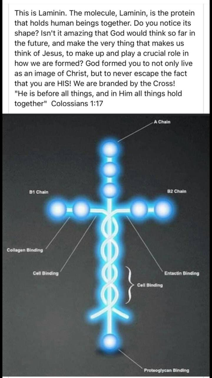
Fig. 3. General structure of laminins. Laminin heterotrimers (Lm’s) exist in three shapes: cruciform, Y- or T-shaped, and rod-shaped. The globular domains of the short arms laminin N-terminal (LN) and laminin four (L4) are separated by rod-like laminin EGF-like repeat (LE) domains. The chains trimerize via the coiled coil domains to form the long arm. The COOH-terminal LG domain is unique to the α chains.
Table 1. Laminin nomenclature and chain composition.
Current name Previous name Chain composition
Laminin 111 Laminin-1 α1β1γ1
Laminin 211 Laminin-2 α2β1γ1
Laminin 121 Laminin-3 α1β2γ1
Laminin 221 Laminin-4 α2β2γ1
Laminin 3A32 Laminin-5A α3Aβ3γ2
Laminin 3B32 Laminin-5B α3Bβ3γ2
Laminin 3A/B11 Laminin-6A/B α3A/Bβ1γ1
Laminin 3A/B21 Laminin-7A/B α3A/Bβ2γ1
Laminin 411 Laminin-8 α4β1γ1
Laminin 421 Laminin-9 α4β2γ1
Laminin 511 Laminin-10 α5β1γ1
Laminin 521 Laminin-11 α5β2γ1
Laminin 213 Laminin-12 α2β1γ3
Laminin 323 Laminin-13 α3β2γ3
Laminin 423 Laminin-14 α4β2γ3
Laminin 523 Laminin-15 α5β2γ3
Laminin 212 α2β1γ2
Laminin 222 α2β2γ2
Laminin 333 α3β3γ3
Laminin 522 α5β2γ2
Extracellular Matrix
Maurice Godfrey, in Asthma and COPD (Second Edition), 2009
Laminin
Laminins are cross-shaped molecules that comprise several different types and are a part of lung development [1, 77–79]. Analysis of the developing lung has shown the expression of some laminin chains as early as 10 weeks of human gestation [80]. Laminins are expressed along the basement membrane, but temporal and spatial expression of different laminin subunits has been documented [25, 81]. Laminin expression may play a role in cell differentiation in the lung as well as other tissues. There is some evidence to suggest that laminin may play a role in alveolar morphogenesis [82]. A laminin–heparan sulfate interaction may be essential to lumen formation and branching morphogenesis [83].
Design considerations when engineering neural tissue from stem cells
Stephanie Willerth, in Engineering Neural Tissue from Stem Cells, 2017
2.1.3 Laminin
Laminin is a protein that shares several properties with fibronectin. For example, it also has a high molecular weight. The laminin protein consists of three subunits, including α, β, and γ chains as shown in Fig. 2. Laminins are also considered to be glycoproteins as they are functionalized with oligosaccharides [3]. In the extracellular matrix, laminin can bind other laminin molecules as well as other proteins like collagen, which helps to reinforce the extracellular matrix structure. Cells can also bind to laminin through the integrin receptors they express on in their cell membranes [15]. These properties make laminins attractive for use as cell culture substrates for both pluripotent stem cells and neural cells [16,17]. Protocols for differentiating stem cells into mature phenotypes will often require culture on 2D laminin substrates. Laminin also plays a key role in the development of the nervous system as axons tend to migrate on this protein, making it attractive for regenerative medicine applications [18]. Finally, laminin is found in the neural stem cell niche, where it contributes its growth factor binding properties to specialized structures known as fractones [19].
Fig. 2
Sign in to download full-size image
Fig. 2. The molecular structure of laminin. It consists of three subunits named α, β, and γ.
Reprinted with permission from Plantman S. Proregenerative properties of ECM molecules. BioMed Res Int 2013;2013.
Neuropathology
Edoardo Malfatti, Norma Beatriz Romero, in Handbook of Clinical Neurology, 2018
Laminin α2
Laminin α2-related dystrophy, MDC1A, also referred to as merosin deficiency, is caused by AR mutations in the LAMA2 gene on chromosome 6q. This form presents at birth with hypotonia, respiratory failure, and contractures. Serum CK is elevated and brain white-matter changes are observed on MRI T2-weighted sequences. Muscle biopsy usually shows prominent fibrosis and fat tissue substitution. Laminins are component of the basal lamina composed by a heterodimer of α, β, and γ chains. Immunohistochemical analysis of laminin α2 in MDC1A shows complete absence, few unlabeled fibers, or reduction of labeling (Fig. 30.10). Two commercial antibodies directed against 300-kDa and 80-kDa fragments of laminin α2 are used in diagnostic routine. Labeling of other laminin chains, such as laminin α5, laminin β1, and γ1, may give additional information. For example, overexpression of laminin α5 is observed in MDC1A mature fibers. Attention should be paid because laminin α5 is highly expressed in immature or regenerating fibers (Sewry et al., 1995). Nerve axons show normal laminin α2 round staining around axon; conversely, they remain unlabeled in MDC1A. Secondary laminin α2 reduction of labeling occurs as a consequence of mutations in other genes responsible for congenital muscular dystrophies.
Sign in to download hi-res image
Fig. 30.10. Fluorescent immunolabeling of laminin α2. (A) Control showing staining of the basal membrane. (B) Reduced expression of laminin α2 in some fibers, and absent expression in others in a molecularly confirmed laminin α2-related dystrophy. (C) Complete absence of staining.
Extracellular Matrix (ECM) Molecules
Jasvir Kaur, Dieter P. Reinhardt, in Stem Cell Biology and Tissue Engineering in Dental Sciences, 2015
3.5.1 Laminins
Laminins are heterotrimeric glycoproteins, composed of α, β, and γ chains. There are 11 genes (five α, three β, and three γ chains) in the human genome that encode these structurally homologous chains, and which form at least 15 trimeric combinations [95]. The nomenclature of laminins is based on the composition of α, β, and γ chains, so that a laminin composed of α2, β1, and γ1 chains is described as laminin 211. Laminins are modular proteins that contain laminin type EGF-like domains, a laminin coiled-coil domain, and carboxyl terminal laminin globular domains that mediate cell adhesion [96]. Laminin α, β, and γ chains trimerize through their coiled-coiled domains to form a cruciform-like structure, the short arms of which are involved in self-assembly and interaction with other basement membrane constituents. Self-polymerization of laminins is a critical step in basement membrane formation, and is dependent on cell membrane interactions with integrins, dystroglycan, the Lutheran glycoprotein, and sulfated glycolipids. These interactions cause actin cytoskeleton rearrangements that in turn have functional consequences on cell behavior. Knockout studies in mice show that most laminin chains are essential for development, with some mice unable to complete gastrulation.
Cross Talk Between Inflammation and Extracellular Matrix Following Myocardial Infarction
Yonggang Ma, … Merry L. Lindsey, in Inflammation in Heart Failure, 2015
4.5.4 Laminins
Laminins, the first ECM glycoproteins detectable in the embryo, are found in basement membrane. Laminins consist of three peptide chains: α, β, and γ. Laminin protein is detected in the infarct area at day 3 post-MI, peaks in concentration at days 7-11, and then returns to baseline levels [69]. The wide existence of laminins throughout the infarct area suggests that they may directly regulate LV repair post-MI [69]. In patients with acute MI, serum laminin level is higher than in patients with stable coronary artery disease and those without coronary artery disease [70]. This report suggests the possibility that serum laminin could be a potential prognostic marker for MI patients.
Molecular and Extracellular Cues in Motor Neuron Specification and Differentiation
R.L. Swetenburg, … L. Karumbaiah, in Molecular and Cellular Therapies for Motor Neuron Diseases, 2017
Laminin
Laminins are large (~800 kDa) ECM-associated glycoproteins thought to be essential for neuronal migration and axonal pathfinding during development. They are heterotrimeric molecules consisting of α, β, and γ chains. These chains present themselves in a variety of different combinations to result in as many as 18 distinct laminin isoforms, a majority of which are present in the nervous system ECM.54 S-laminin is a homolog of the B1 subunit of laminin. It is known to facilitate MN adhesion, and plays an important role in the formation of neuromuscular junctions. In the developing CNS, S-laminin and laminin are both found to be present in the subplate in the cerebral cortex.55 This situation changes dramatically in the adult CNS, which is marked by the disappearance of S-laminin, and the restricted expression of laminin to the basement membrane, where it is found in close association with collagen and heparan sulfate proteoglycans (HSPGs).51
Laminins are important constituents of the PNS basement membrane found surrounding Schwann cells. As integral adhesive and growth-promoting constituents of the basement membrane, they are believed to organize together the meshwork of structural proteins to form the substratum upon which neuronal migration and axonal pathfinding can take place.56 The diversity of laminin isoforms can be advantageous in regulating the selective attachment of MNs, which is reportedly mediated by the S-laminin-specific LRE (leucine–arginine–glutamic acid) amino acid sequence.57
Integrin Regulation of the Lung Epithelium
Erin Plosa, Roy Zent, in Lung Epithelial Biology in the Pathogenesis of Pulmonary Disease, 2017
5.1.2.1 Laminins
Laminins, along with collagen IV, provide substantial structural support for the lung and are the primary components of the lung basement membrane. Laminins are heterotrimeric proteins, composed of one α, one β, and one γ chain, and have 16 confirmed or predicted human isoforms formed from five α chains, three β chains, and three γ chains [30]. The fetal lung basement membrane contains laminins with all five possible α chains. In contrast, the adult lung epithelium restricts α chain expression to laminin α3 and α5 [31–33].
Targeted laminin subunit deletions or inhibition demonstrate the requirement for laminins in lung development [34,35]. Inhibition of laminin α1 in fetal lung explants results in branching morphogenesis defects and disrupted epithelial polarity [36]. Global deletion of laminin α5 results in fetal loss in mid-late gestation associated with impaired lobar septation of the lung [37]. Inducible lung epithelial specific deletion of laminin α5 demonstrated additional roles in lung development. These mice died from respiratory failure shortly after birth. However, histological examination of their lungs in late fetal lung development revealed epithelial differentiation defects, dilated distal airspaces, increased apoptosis, and decreased proliferation in whole lung sections [38].
The role of laminin 332 during lung development has been investigated using global and tissue specific murine deletions, fetal lung explants, and in-vitro studies. The expression of laminin 332 localizes exclusively to the basal surface of epithelial cells in the fetal lung basement membrane. In contrast to laminin α1 and α5, the γ2 chain (specific to laminin 322) is not required for branching morpho genesis or fetal lung epithelial cell differentiation. Histological analysis of lungs from laminin γ2−/− mice demonstrated disorganized hemidesmosomes in the tracheal epithelium, yet these mice retained normal epithelial integrity and lung function [39]. The role of laminin γ2 in the adult lung was not addressed with this genetic approach, as severe skin defects in the laminin γ2−/− mice resulted in perinatal lethality from malnutrition [40]. Laminin 322 is also expressed in the adult lung basement membrane. In this context, lung epithelial specific deletion of laminin α3 demonstrated a role for this laminin isoform in the development of pulmonary fibrosis [41]. These mice lack a developmental phenotype, but exhibit increased mortality, inflammation, and fibrosis following intratracheal bleomycin challenge.
Axonal Pathfinding: Extracellular Matrix Role
P. Letourneau, in Encyclopedia of Neuroscience, 2009
Laminins
Laminins are large adhesive glycoproteins (MW 1 000 000 Da) that consist of heterotrimers of alpha, beta, and gamma chains. Ten laminin chains are known, forming 11 known heterotrimers with widely varied expression throughout different tissues. The laminins present several domains that mediate laminin binding to several cell surface receptors and to other ECM components. The most common laminins are typically present in basal laminae, an ultrastructural ECM layer associated with epithelia, muscle cells, Schwann cells, and glia. Laminin-1, which has been studied the most, promotes axonal growth in vitro from virtually every type of neuron, indicating that laminins have broad roles in promoting growth cone migration. Examples of growth cone migration along basal laminae include growth cones of Rohon–Beard neurons in Xenopus, growth cones of retinal ganglion cells in the retina and optic nerve, and pioneer axons in the grasshopper limb bud. However, in addition to basal laminae, laminin is transiently expressed in the loose cellular environments of developing tissues, including the nervous system, on cell surfaces and associated with sparse ECM fibers. The growth cones that pioneer pathways, such as the corticofugal pathway of the neocortex or the medial longitudinal fasciculus from the brain into the spinal cord, migrate within loose extracellular spaces in the wall of the immature central nervous system (CNS), where the cells are labeled in a punctate manner by laminin antibodies. The expression of laminin on these cells is transient, and eventually laminin immunoreactivity is restricted to the basal lamina at the outer boundary of the CNS wall. In the developing peripheral nervous system (PNS), laminin is expressed in basal laminae and at early stages in the mesenchyme through which motor and sensory axons extend. Schwann cells express abundant laminin, forming the basal laminae that enclose axon–Schwann cell units. This punctate cellular expression of laminin diminishes during development, although laminin remains present in basal laminae.
In view of the wide distribution of laminins and the ability of laminin-1 to promote robust axonal growth from many neuronal types, it is thought that laminins function permissively, providing adhesion that is required for growth cone migration, but not in an instructive manner to influence pathfinding decisions. Laminins and other ECM molecules may broadly promote growth cone migration along a pathway, whose boundaries are defined not by the absence of adhesive ECM molecules but, rather, by the expression in adjacent tissues of negative guidance cues, such as slits or semaphorins. This ‘surround repulsion’ occurs in both developing CNS and PNS. Several mutational studies have reported specific errors in pathfinding when a laminin is absent or blocked. Laminin function is essential for growth cone turning in the grasshopper limb bud, and zebra fish with mutations in the laminin-alpha-1 chain exhibit multiple axon guidance defects throughout the CNS, but not in every location. These results suggest that laminin-mediated adhesion is essential for growth cone navigation in at least some instances.
https://www.sciencedirect.com/topics/neuroscience/laminin


